
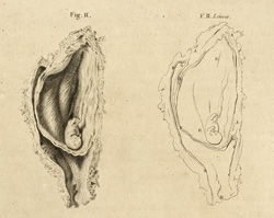
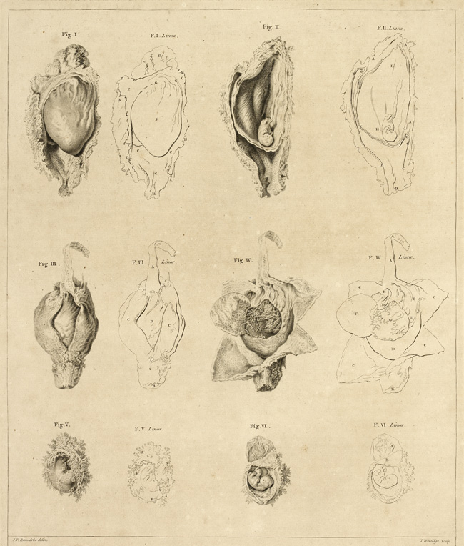
Anatomy of the gravid uterus. The best-known atlas of pregnancy is The Anatomy of the Human Gravid Uterus (1774) by William Hunter (1718–83), a successful surgeon, man-midwife and anatomy teacher. A largely French team engraved the 34 plates, mostly after drawings by the London medical artist Jan van Rymsdyk. The naturalistic approach of British medical illustration in this period chimed with Hunter’s preference for minimal intervention. The most famous and shocking figures show the child lodged in the trunk between the mother’s sawn-off limbs. But though Hunter focused on the uterus, he also pictured embryos from what he estimated as three weeks to almost full term.
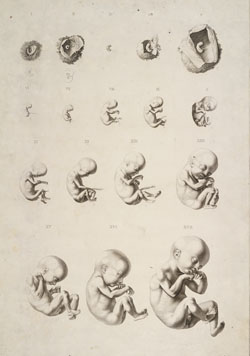
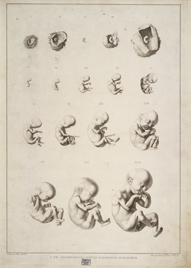
Soemmerring’s Images of human embryos, 1799
In 1799 a German anatomist produced what is generally considered the first connected series of images to show the development of human embryos.
In the late 1700s governments and medicine focused intensely on pregnancy because a healthy and numerous population was now seen as essential to a well-ordered, competitive state. The punishment and prevention of abortion and infanticide were hotly debated in legal and medical circles. New lying-in hospitals provided care, food and shelter for poor, unmarried women. They also trained midwives and the man-midwives, later called obstetricians, who were taking over childbirth among the upper classes. These institutions increased anatomists’ access to embryonic and fetal specimens.
Anatomists explored the pregnant uterus and its contents. The London man-midwife William Hunter concentrated on the uterus, but the German anatomist Samuel Thomas Soemmerring focused on the embryo. In true Enlightenment spirit, his ‘images of human embryos’ sought to reveal the mystery hidden in the dark womb. They show not just growth but change in shape.