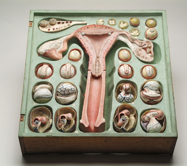
‘Eggology’ in papier mâché.
The now visible egg and other embryological discoveries
attracted model-makers’ attention. The most commercially successful
was Dr Louis Auzoux, whose factory furnished educational institutions
and individuals in France and abroad with innovative models
in papier mâché, a material more robust than wax, and so more
suitable for teaching. Many of Auzoux’s models could be taken
apart and put together again. This 21-piece ‘ovologie’ represents
the human female reproductive system and vertebrate embryonic
development from ovulation to the ‘formation of the embryo’.
The earliest known reference to this series comes from the sales
catalogue attached to the textbook, Leçons élémentaires
d’anatomie et de physiologie humaine et comparée (1858,
pp. 350–1), but the pictured set was produced after Auzoux’s
death in 1880.
41 x 43 x 11 cm.
Museum Boerhaave, Leiden.
The mammalian egg. Since
de Graaf, the large follicle in
the ovary was regarded as the egg, but many scholars doubted his results.
They wondered, among other things, how such a big object could pass through
the narrow tubes. In the mid-1820s, Karl Ernst von Baer
was certain that the egg must originate in the ovary but that it was not
the follicle de Graaf had observed. He had his teacher and colleague Karl
Burdach give him a house bitch that had gone into heat a few days earlier.
Seeing only follicles in the ovaries, Von Baer was about to give up, when
with the unaided eye he noticed ‘a yellow spot’ in some of them. Opening
one, he lifted the spot into a watch glass for microscopy. He had hoped
so much for this sight, but when it finally came he recoiled ‘as if struck
by lightning, … afraid I had been deluded by a phantom’. The discovery of
a tiny elementary structure, from which a whole organism developed, was
an important inspiration for the later cell theory.
Cells and disciplines
Microscopy led to the discovery
of cells and embryology was institutionalized in medical courses.
Researchers in German institutes of anatomy and physiology followed
Pander and von Baer. After the discovery of
the true mammalian ovum, a more general cell theory was developed
from the late 1830s. In the early 1850s, Robert Remak, whose Jewish faith
hindered his career at the University of Berlin, argued that all cells arise
from pre-existing cells, from the egg via the germ layers to the tissues.
He also taught that in vertebrates each layer gives rise to particular cell
types, for example, liver from endoderm, muscle from mesoderm and nerve
from ectoderm.
By the 1850s special anatomical courses for medical students were
embryology’s main base. Developmental approaches were very important,
but the science achieved only limited institutional independence. A new,
physics-oriented physiology reckoned embryos intractable with physico-chemical
methods and abandoned the subject to anatomy and the newly independent zoology
institutes. Few human specimens from the first month and none from the first
two weeks were known, but by teaching that vertebrate development follows
a common pattern, comparative embryology licensed the use as surrogates
of frog, chick and domestic mammals.
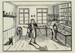

An optician’s workshop. The foundation of the
University of Berlin in 1810 and the expansion of the
medical sciences stimulated the crafts of instrument-makers
and opticians, whose numbers in the Prussian capital
dramatically increased. The microscope was highly sought-after,
especially following the invention of achromatic technology
in the 1830s. While earlier images had been surrounded
by coloured fringes resulting from the refraction of
natural light, now scientists saw vividly and sharply.
The expensive new microscopes were initially purchased
only for the private laboratories of professors and
by wealthy medical students. Later, laboratory training
was extended to every student and cheaper microscopes
became standard equipment for all.
Woodcut titled ‘Mechaniker-Werkstatt’ (1832) from
Franz Maria Feldhaus, Carl Bamberg. Ein Rückblick
auf sein Wirken und auf die Feinmechanik, Berlin:
Askania-Werke, 1929, fig. 13, p. 23. 24 x 17 cm.
Niedersächsische Staats- und Universitätsbibliothek,
Göttingen.
An optician’s workshop, 1832
|
|
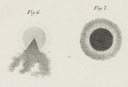
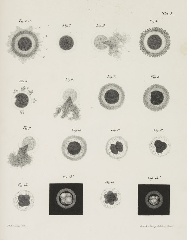
Cleaving dog embryos. Inspired by
von Baer, in the early 1840s
the Giessen anatomist Theodor Bischoff authored pioneering
studies of early mammalian development, first of the
rabbit and then of the dog. Even for these relatively
accessible species, the construction of developmental
series was severely limited by what he could collect
from animals he bred and bought. These lithographs show
cleavage in dog eggs taken from the ovary (figs 1–6)
and the Fallopian tubes (figs 7–14). Three have been
opened with a needle to release the yolk and with it
the ‘germinal vesicle’ (the modern nucleus). Figures
13* and 14* are reversed, white on black, to mimic the
effect of reflected-light microscopy. Bischoff was still
cautious about interpreting cleavage as a process of
cell division, but these and their rabbit counterparts
were soon reproduced as wood-engravings
in textbooks and more popular works, and also as wax
models.
Lithograph by A. Schütter after Bischoff’s drawings,
printed by Henry & Cohen, from Th. Ludw. Wilh. Bischoff,
Entwicklungsgeschichte des Hunde-Eies, Braunschweig:
Friedrich Vieweg und Sohn, 1845, plate I, 27 x 21 cm.
By permission of the Syndics of Cambridge University
Library.
Lithograph of cleaving dog embryos, 1845
|