Visual aids
Embryology classes relied unusually heavily on a battery of carefully prepared images to show students what they were supposed to see.
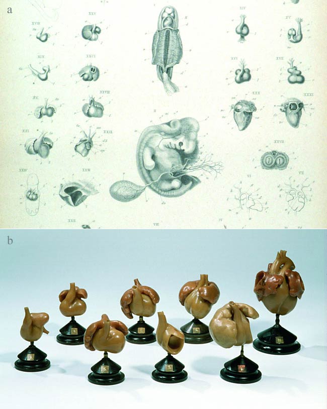
Anatomists had long used visual aids to convert the messy experience of dissected cadavers into well-ordered mental maps. Yet embryology posed special challenges. While adult cadavers were large, robust and relatively abundant, embryos, especially early human ones, were tiny, fragile and scarce. Each also represented just one moment in a complicated set of transformations. So by the mid-1800s lecture-demonstrations and practical classes supplemented prepared specimens with highly magnified wall charts, blackboard drawings, large-format atlases and increasingly highly illustrated textbooks. Wax models made complex three-dimensional structures much easier to grasp.
Success was mixed. Keen students and lay listeners were extremely excited to see embryos for the first time, but as the courses became routine many medics found the complicated bendings and foldings of cell layers, and the abstruse terminology, difficult and dull.
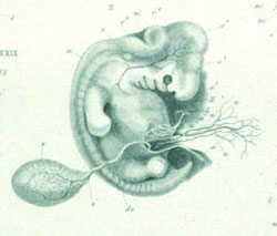
Engravings and models of heart development, c.1858 |
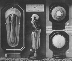
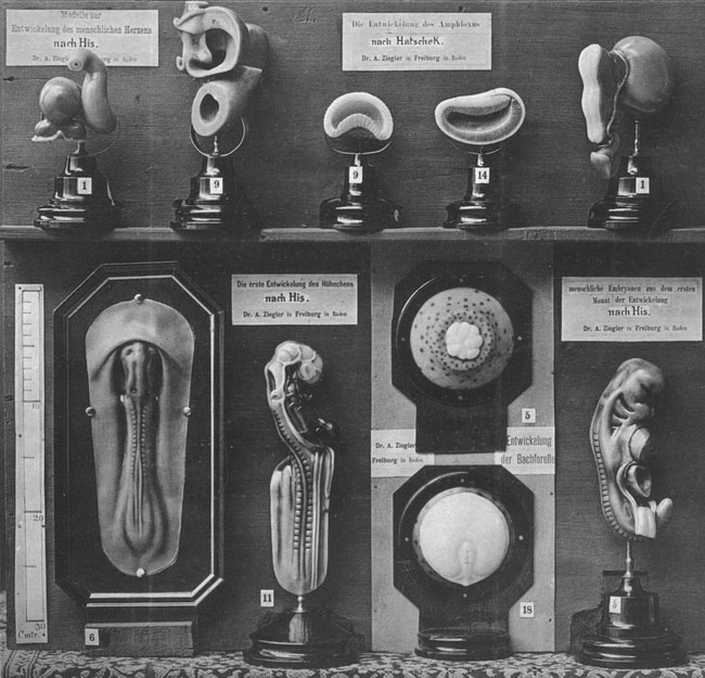
Display by the ‘plastic publisher’ Adolf Ziegler, c.1886 |