Anatomy of human embryos
An innovative collecting strategy plus new modelling techniques produced a much more detailed and vivid understanding of early human development.
Wilhelm His amassed several dozen embryos from the first two months by organizing a supply network, especially of gynæcologists. Human embryology was still limited above all by the difficulty of collecting material. To encourage donations, mostly from spontaneous abortions, he railed against clinicians who wasted ‘precious objects’ by leaving them on the shelf or analysing them incompetently. But he rewarded those who gave up their ‘treasures’ by naming the specimens after the donors—the gynæcologists, that is, not the women from whose bodies they came.
His took the specimens through a complex sequence of image-making operations. First he dissected away the egg membranes, and hence the remaining evidence of connection to the pregnant woman, and drew and photographed the embryos he found inside. Then he put these under ‘the guillotine’ of the section-cutter, or microtome, and for the first time converted them completely into slices on microscope slides. This gave unprecedented access to internal structure. But the crucial step was modelling, which, as he put it, ‘gave’ the sectioned embryos ‘body’.
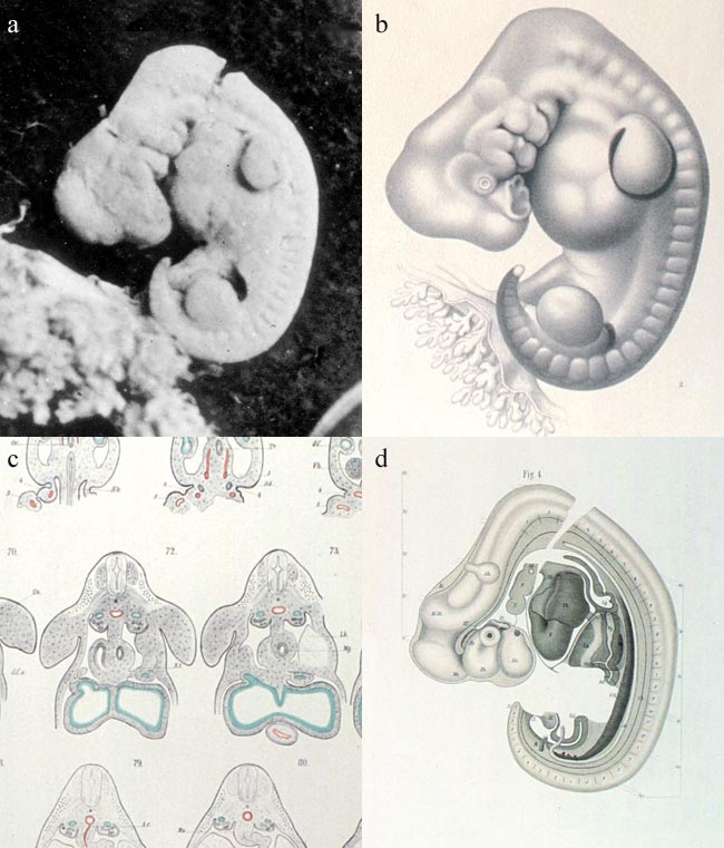
Photograph and drawings of embryo A, 1880–5 |
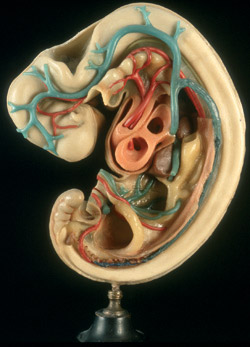
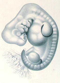
Models of embryo A, c.1888 |