Making obstetric ultrasound
In the 1950s, ultrasound promised a safe window into the womb.
Ultrasound, a technology that converts reflected sound waves into electrical impulses, was first developed to locate submarines in World War I. By the 1940s, ultrasound was used in engineering to detect flaws in metal structure and, by the 1950s, in clinical medicine for diagnostics as well as, at high energy, for therapy. Around 1950, the Denver radiologist Douglas Howry immersed patients in water to improve the quality of cross-sectional ultrasound scans; in Minnesota, the surgeon John Wild attempted to construct a device for quickly distinguishing normal from cancerous tissue.
In 1955, in the shipbuilding and industrial centre of Glasgow, the up-and-coming gynæcologist-obstetrician Ian Donald borrowed the ultrasound equipment from a local firm to differentiate abdominal masses indistinguishable by palpation. Sceptics reckoned the pictures unintelligible; others suggested that obstetricians experience of blind palpation trained them to assemble the pieces of a puzzle into a mental image. On the whole, by the late 1960s the diagnostic potential of obstetric ultrasound had become widely acknowledged.
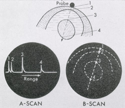
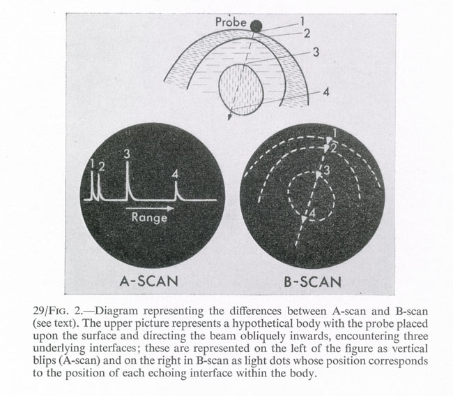
Technologies of ultrasound scanning, 1979 |
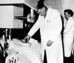
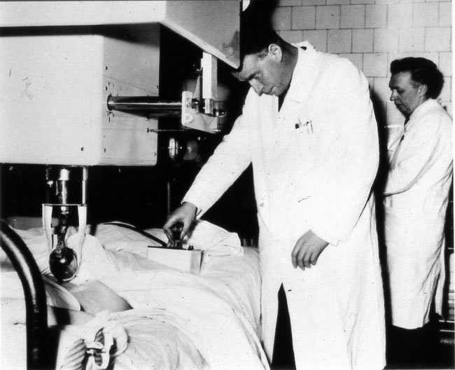
Ian Donald and John MacVicar examining a patient, 1960 |