
Uses of ultrasound
Ultrasound was quickly accepted as a diagnostic tool, but the modes of use, frequency of examination and norms of safety and fetal development are still not fully standardized.
Between the 1950s and 1970s, physicians, physicists and engineers debated whether the scan should show a morphological image, akin to X-rays, or a graphical representation of deviations from the norm, like an electrocardiograph. In obstetrics the morphological orientation won, but different countries decided differently about frequency of use and indications for scanning, depending on whether ultrasound was framed as a diagnostic aid solely for pregnancies at risk, or a routine screening device for all.
Uses expanded beyond diagnosis. Around 1970, psychologists proposed that ultrasound could help the concerned mother bond with the future child. Yet that bonding would have to be mediated, because at the time no untrained eye could interpret the scans. Professionals did this instead: obstetricians, midwives and specialized sonographers. Gradually, easier-to-read sonograms came to occupy a symbolic place in late twentieth-century popular culture. Many parents first visual encounter with their child was no longer at birth but seeing the gestational saca bright ring around a clear centre, characteristic of early pregnancyor, even more powerfully, the embryonic heart beating on an ultrasound screen.

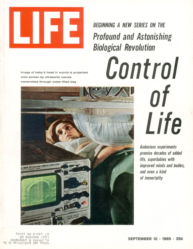
‘Control of life’ in Life, 1965 |
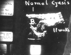
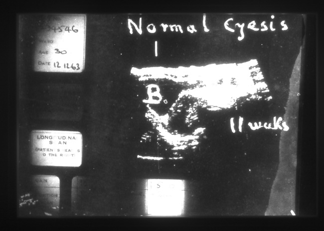
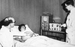
Interpreting ultrasound scans, 1963 |