The Boston egg hunt
In the late 1930s the Carnegie Department set out to recover embryos younger than 14 days from gynæcological operations.
Before a woman missed her period and the new hormonal pregnancy tests could turn positive, embryos remained undetectable. So in 1938, John Rock, a Boston gynæcologist, human fertility researcher and practising Catholic, teamed up with a pathologist with Carnegie research experience on fertilization in monkeys, Arthur T. Hertig, and technician Miriam Menkin. She became Rocks long-term collaborator as they looked for the earliest products of conception in surgically removed Fallopian tubes and uteri.
Yet spotting such an early human embryo was very hard, especially since no one had seen one before. Hertig reported that we looked at an enormous amount of eggs that werent eggs, just cellular debris, because
the art of actually recognizing a human egg hadnt been mastered. In late 1938, he described the first complete fertilized egg he saw under his microscope as a glistening bead, like a pearl of tapioca. The 12-day Harvard egg was examined, measured and then transported to Baltimore. The 34 fertilized eggs from 211 unknowingly pregnant women, collected by Hertig and Rock over the course of 14 years, came to represent the first 17 days of life.
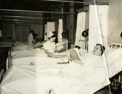
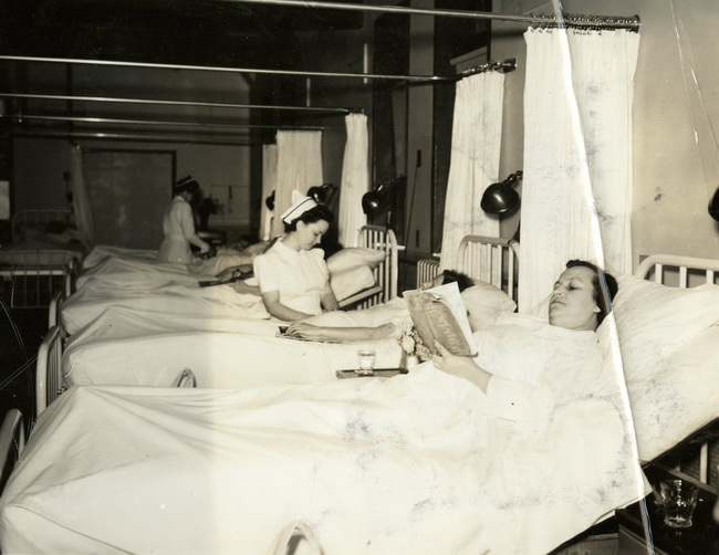
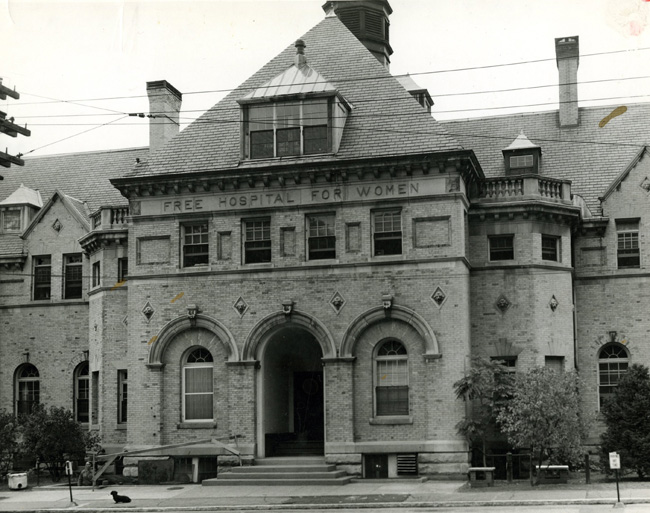
The Free Hospital for Women. Between 1926 and 1955 this prestigious institution
housed John Rocks Fertility and Endocrine Clinic and was the source of many early embryos in the Carnegie collection.
The hospital had been founded in 1875 to provide care for largely Roman Catholic patients from lower socio-economic groups.
The building was erected in 18945 from yellow brick and in the style of a French estate house, on a riverside glen in Brookline,
a town bordering Boston. The hospital was a research facility for Harvard Medical School, so all women, clinic as well as private
patients accommodated in the Parkway wing, were research subjects as well as patients.
Photograph, c.1950, credited to Robert J. Keller, Countway Library of Medicine, Harvard University.
The Free Hospital for Women, associated with the Harvard Medical School, c.1950
| |
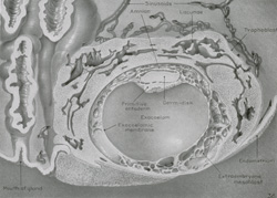
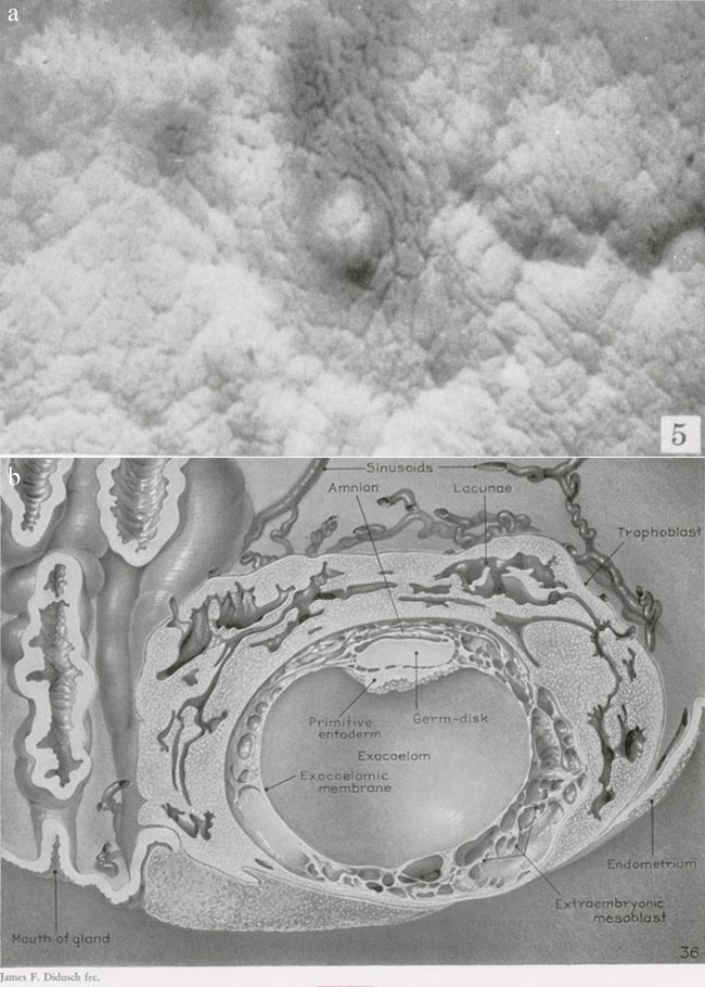
The Harvard egg. This egg came from a 34-year-old white woman who in 14 years of marriage gave
birth to nine live children and one six-months fetus. She had long suffered pelvic discomfort and general malaise that worsened
after her last delivery in March 1938. Her abdomen was found to be free from masses and pain, but her cervix, lacerated and
frequently bleeding especially after intercourse, showed evidence of chronic inflammation and malignant changes. On 28 November
1938, her uterus and right ovary and tube were removed. (a) The photograph shows the implantation site in the uterine mucous
lining, while (b) the drawing based on a photomicrograph shows parts of the embryo.
(a) Photograph by Chester Reather (figure 5 on plate 1) and (b) drawing by James Didusch (plate 8) from Arthur T. Hertig and John Rock, Two human ova of the previllous stage, having an ovulation age of about eleven and twelve days respectively, Contributions to Embryology 29, 1941, 12756. 28.5 x 22.5 cm.
Carnegie Institution of Washington.
Drawing of the Harvard egg, 1941
|