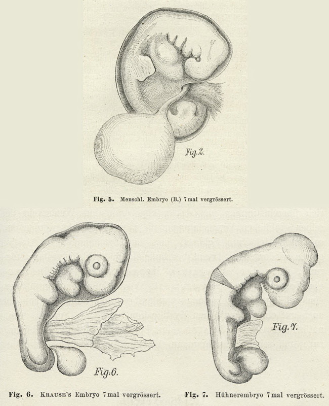
Comparing chick and human embryos.
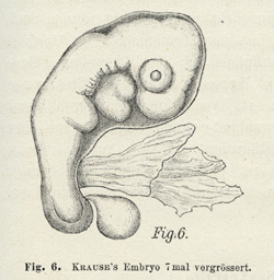
Drawings by Wilhelm His comparing his human embryo (fig. 5),
Krause’s embryo (fig. 6), and a chick embryo (fig. 7). The disputed
part was the allantois, a structure known in birds and some
mammals to form a bridge between the embryo proper and the chorion
that was crucial to embryonic nutrition. Krause’s embryo featured
what he claimed was a prominent free allantois (the vesicle
near the posterior end), but His argued that in humans the allantois
was never visible in this form and reinterpreted the specimen
as that of bird. Either a bottle had been mislabelled or Krause
had been deceived.
Wood-engravings from Wilhelm His, Anatomie menschlicher
Embryonen, part 1: Embryonen des ersten Monates,
Leipzig: Vogel, 1880, figs 5–7, pp. 70–1. Drawing of Embryo
B 6.6 cm tall.
Excluding an embryo. The story of the
embryo it took His most effort to exclude shows how open human embryology
still was when he began work. In 1875 an anatomy professor, Wilhelm Krause,
had described a human embryo as supporting Haeckel against His. For five
years embryologists debated the possibility that the embryo was abnormal,
but in 1879 His announced that it was not human at all, but a chick. Even
then, Krause fought on and others continued to offer alternative explanations.
The matter was decided only in 1882, when anatomists gathered to view the
famous object. They declared it a bird, but that it was a chick they could
not say; it might just as well have been a duck, a goose—or a turkey.
Setting standards
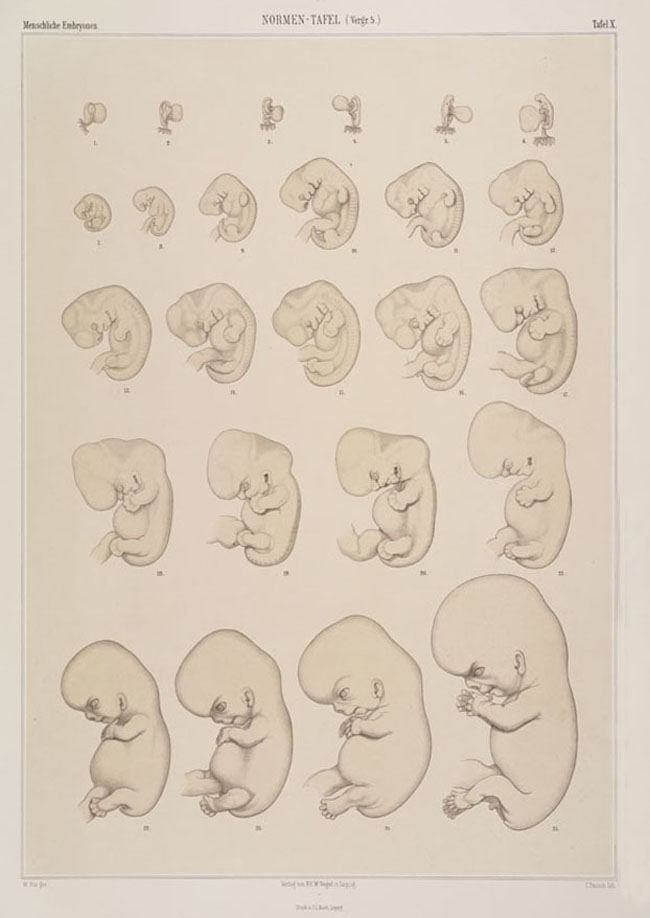
Wilhelm His invented a new kind of visual standard, a ‘normal plate’.
His arranged specimens in developmental order and selected appropriate
representatives. This was far from trivial. He could not simply sort
the embryos by time from fertilization, since this was not reliably known,
so instead set up a special measure of length. For the variously shaped
earliest specimens, he fell back on his judgement of overall form. Selecting
normal specimens from material that, by its very origin in abortions, was
suspected of abnormality was even harder. His could only compare his own
and published descriptions among themselves as he
drew and redrew, excluding venerable
specimens and some undistinguished recent offerings from gynæcologists.
Though His began by setting up ‘stages’ of human development, he soon
abandoned this system in favour of a series of ‘norms’. These were not,
as for Soemmerring, the most perfect, but merely
individuals he took to be characteristic, well preserved and well described.
So though His faced fundamentally the same problems, he solved them in significantly
differently ways. He called the resulting seriation a Normentafel
or ‘normal plate’.
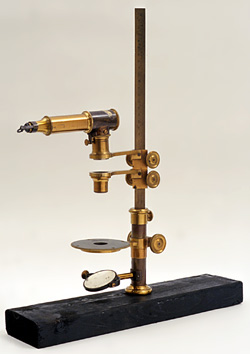
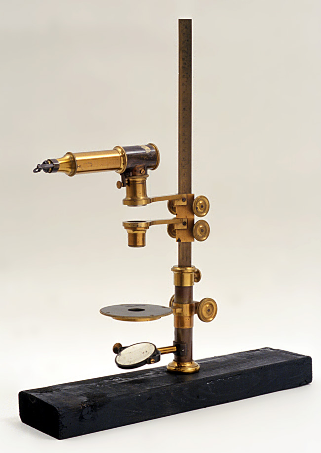
A drawing apparatus or ‘embryograph’. For His,
exact drawings of whole embryos and sections were the
foundation of all serious work. Committed to mechanical
drawing aids, he found the standard combination of microscope
and camera lucida unsuitable for the low magnifications
(5–20 x) that embryological specimens required. So he
replaced the microscope with lenses freely movable on
a graduated retort stand. From the early 1880s Edmund
Hartnack of Paris and Potsdam sold the apparatus as
an ‘embryograph’. We see a drawing prism (top), objective
lens, stage and mirror (bottom); the image was projected
onto a drawing table. The embryograph was widely used,
but Haeckel, defending himself against
accusations of tendentious drawing,
mocked His for reducing the morphologist to an automaton-like
‘embryographer’.
Embryograph, total height of c.45 cm, acquired
from a Dr M. Abraham in 1942.
Historical Collections, National Museum of Health
and Medicine, Armed Forces Institute of Pathology, Washington
D.C.
A drawing apparatus or ‘embryograph’,
late 1800s
|
|
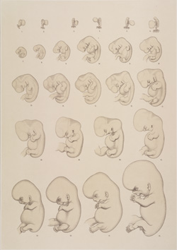
Normal plate by Wilhelm His. His had his artist
draw embryos from about the end of the second week to
the end of the second month. The plate almost creates
the impression of a single embryo at a succession of
stages, but in fact brought together specimens from
diverse medical encounters. The ninth figure shows
embryo A.
Lithograph by C. Pausch from Wilhelm His, Anatomie
menschlicher Embryonen, part 3: Zur Geschichte
der Organe, Leipzig: Vogel, 1885, plate X. Border
46 x 33 cm.
By permission of the Syndics of Cambridge University
Library.
Normal plate by Wilhelm His, 1885
|