Publishing in wax and print
Models became central to research as well as teaching in human embryology.
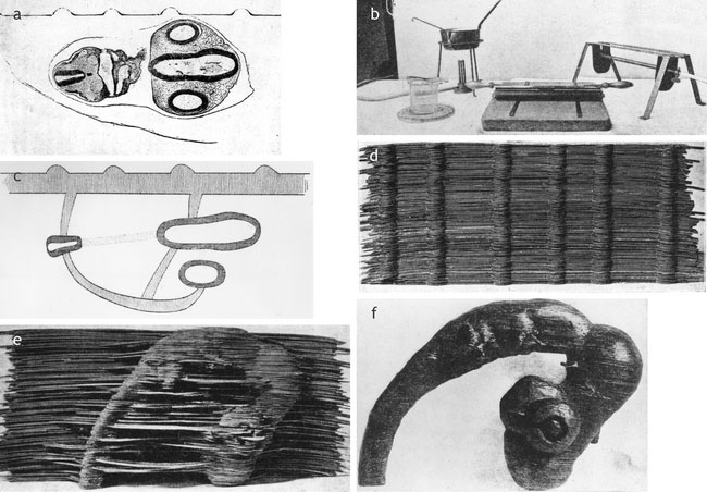
The late nineteenth century was a great age of print, and textbooks, handbooks and journals thrived. Academics tended to look down on other visual aids. But His argued that to grasp complex embryonic forms scientists had to reconstruct models from the sections through which they explored interior structure. And to communicate this understanding they had to make the models available to others. To this end he taught human and comparative vertebrate embryologists to publish in wax as well as print.
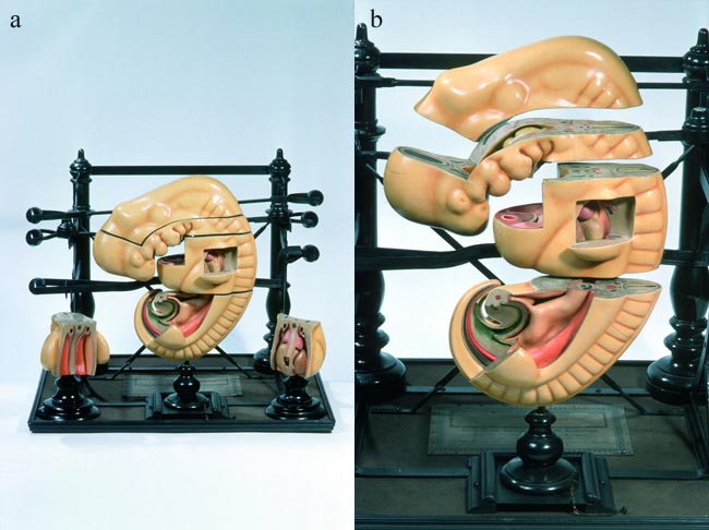
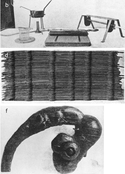
Scientists made and described models, then sent the manuscripts to editors or conventional print publishers and the original models to a self-styled ‘plastic publisher’, usually Adolf Ziegler’s son Friedrich in Freiburg in Baden. Friedrich Ziegler turned the raw originals into effective visual aids that he reproduced and sold worldwide. The models were indispensable in research as well as teaching: journal articles depicted the reconstructions. During the 1880s a wax-plate method replaced His’s freehand modelling, allowing much larger, more detailed and fragmented models.

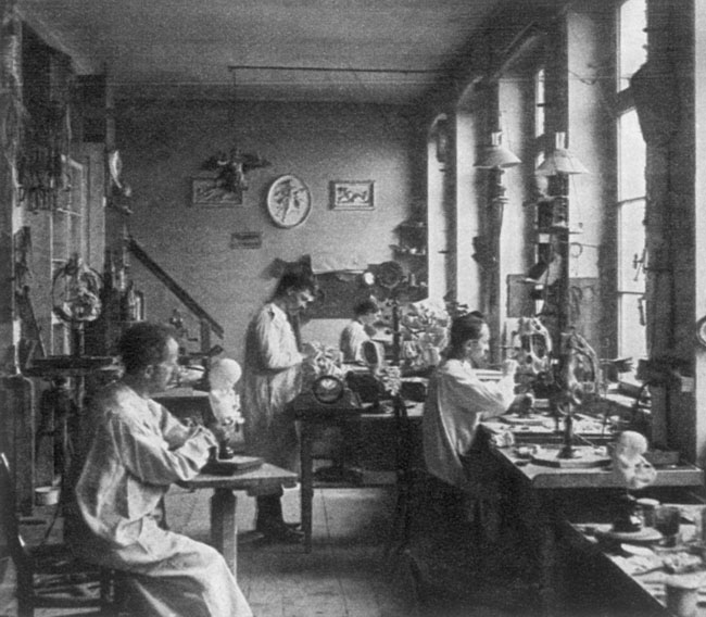
Friedrich Ziegler modelling, c.1912 |
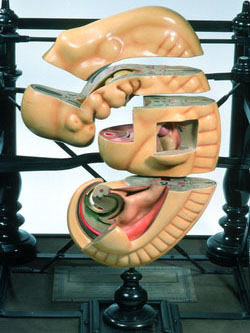
A wax embryo on the rack, c.1902 |